Unit 16: The Peripheral Nervous System
Unit Outline
Part 1: The Peripheral Nervous System
Part 2: The Somatic Nervous System
Part 3: The Autonomic Nervous System
Practice Questions
Learning Outcomes
At the end of this unit, you should be able to:
I. Describe the components of the peripheral nervous system.
II. Describe the components of a reflex arc and explain how a reflex arc works.
III. Describe the function of the autonomic nervous system (ANS) and compare the specific functions of the parasympathetic and sympathetic divisions of the ANS.
IV. Explain how agonists and antagonists of cholinergic receptors were discovered, and how they function at nicotinic and muscarinic receptors.
Part 1: The Peripheral Nervous System
The peripheral nervous system is not as contained as the central nervous system because it is defined as everything that is not the central nervous system. Some peripheral structures are incorporated into the other organs of the body. In describing the anatomy of the peripheral nervous system, it is necessary to describe the common structures, the nerves and the ganglia, as they are found in various parts of the body. Many of the neural structures that are incorporated into other organs are features of the digestive system; these structures are known as the enteric nervous system and are a special subset of the peripheral nervous system.
Ganglia
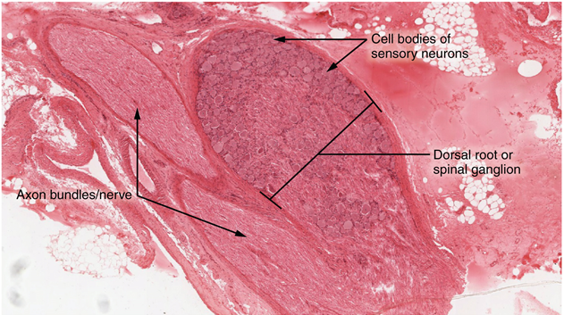
A ganglion is a group of neuron cell bodies in the periphery. Ganglia can be categorized, for the most part, as either sensory ganglia or autonomic ganglia, referring to their primary functions. The most common type of sensory ganglion is a dorsal root ganglion. These ganglia are the cell bodies of neurons with axons that are sensory endings in the periphery, such as in the skin, and that extend into the central nervous system through the dorsal nerve root.
The other major category of ganglia, those of the autonomic nervous system, will be examined later in this chapter.
Nerves
Bundles of axons in the peripheral nervous system are referred to as nerves (Figure 2). These structures in the periphery are different than the central counterpart, called a tract. Nerves are composed of more than just nervous tissue. They have connective tissues invested in their structure, as well as blood vessels supplying the tissues with nourishment. Nerves are associated with the region of the central nervous system to which they are connected, either as cranial nerves (12 pairs) connected to the brain or spinal nerves (31 pairs) connected to the spinal cord.
The cranial nerves are primarily responsible for the sensory and motor functions of the head and neck, although one of these nerves, the vagus, targets organs in the thoracic and abdominal cavities as part of the parasympathetic nervous system. They can be classified as sensory nerves, motor nerves, or a combination of both, meaning that the axons in these nerves originate out of sensory ganglia external to the cranium or motor nuclei within the brain stem.
All of the spinal nerves are combined sensory and motor axons that separate into two nerve roots. The sensory axons enter the spinal cord as the dorsal nerve root. The motor fibres, both somatic and autonomic, emerge as the ventral nerve root. The dorsal root ganglion for each nerve is an enlargement of the spinal nerve.
Part 2: The Somatic Nervous System
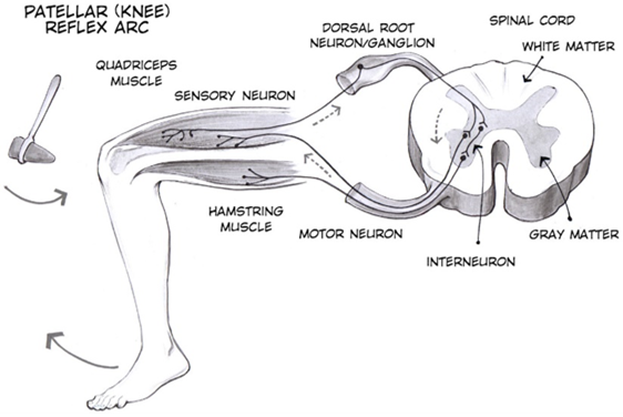
The somatic nervous system is traditionally considered a division within the peripheral nervous system. However, this misses an important point: somatic refers to a functional division, whereas peripheral refers to an anatomic division. The somatic nervous system is responsible for our conscious perception of the environment and for our voluntary responses to that perception by means of skeletal muscles. Peripheral sensory neurons receive input from environmental stimuli, but the neurons that produce motor responses originate in the central nervous system. The distinction between the structures of the peripheral and central nervous systems and the functions of the somatic and autonomic systems can most easily be demonstrated through a simple reflex, an automatic response that the nervous system produces in response to specific stimuli. The neurons and neural pathways responsible for a reflex action constitute the reflex arc. One of the simplest reflex acts is the stretch reflex, by which the nervous system responds to the stretching of a muscle (the stimulus) with contraction of that same muscle (the response). This response protects the muscle from over-stretching, but more importantly, it has a crucial role in maintaining posture and balance. The patellar reflex (or knee-jerk reflex) is an example of stretch reflex and it occurs through the following steps (Figure 2):
- Tapping of the patellar tendon with a hammer causes the stretching of muscle fibres in the quadriceps muscle, which stimulates sensory neurons innervating those fibres.
- In the sensory neuron, a nerve impulse (action potential) is generated, which travels along the sensory nerve fibre from the muscle, through the dorsal root ganglion, to the spinal cord.
- The sensory neuron stimulates a motor neuron in the ventral horn of the spinal cord.
- That motor neuron sends a nerve impulse (action potential) along its axon.
- This impulse reaches the quadriceps muscle, causing its contraction and the extension of the leg (a kick).
The sensory neuron can also activate an interneuron (e.g., Figure 3), which inhibits the motor neuron responsible for the contraction of the antagonistic muscle to quadriceps (i.e. hamstring).
Another example of a simple spinal reflex is the withdrawal reflex, which occurs, for example, when you touch a hot stove and pull your hand away. This reflex occurs through a similar sequence of steps:
- Sensory receptors in the skin sense extreme temperature and the early signs of tissue damage.
- In a sensory neuron, a nerve impulse (action potential) is generated, which travels along the sensory nerve fibre from the skin, through the dorsal root ganglion, to the spinal cord.
- The sensory neuron stimulates a motor neuron in the ventral horn motor of the spinal cord.
- That motor neuron sends a nerve impulse (action potential) along its axon.
- This impulse reaches the biceps brachii, causing contraction of the muscle and flexion of the forearm at the elbow to withdraw the hand from the hot stove.
The basic withdrawal reflex includes sensory input (the painful stimulus), central processing (the synapse in the spinal cord), and motor output (activation of a ventral motor neuron that causes contraction of the biceps brachii). As seen for the patellar reflex, the withdrawal reflex can also include inhibition of the antagonistic muscle (triceps brachii in our example). Another possible motor output of the withdrawal reflex is cross extension: counterbalancing movement on the other side of the body by stimulation of the extensor muscles in the contralateral limb.
The somatic nervous system also controls voluntary movement and more complex motor functions. For example, reading of this text starts with visual sensory input to the retina, which then projects to the thalamus, and on to the cerebral cortex. A sequence of regions of the cerebral cortex process the visual information, starting in the primary visual cortex of the occipital lobe, and resulting in the conscious perception of these letters. Subsequent cognitive processing results in understanding of the content. As you continue reading, regions of the cerebral cortex in the frontal lobe plan how to move the eyes to follow the lines of text. The output from the cortex causes activity in motor neurons in the brain stem that cause movement of the extraocular muscles through the third, fourth, and sixth cranial nerves. This example also includes sensory input (the retinal projection to the thalamus), central processing (the thalamus and subsequent cortical activity), and motor output (activation of neurons in the brain stem that lead to coordinated contraction of extraocular muscles).
Part 3: The Autonomic Nervous System
The autonomic nervous system is often associated with the “fight-or-flight response,” which refers to the preparation of the body to either run away from a threat or to stand and fight in the face of that threat. To suggest what this means, consider the (very unlikely) situation of seeing a lioness hunting out on the savannah. Though this is not a common threat that humans deal with in the modern world, it represents the type of environment in which the human species thrived and adapted. The spread of humans around the world to the present state of the modern age occurred much more quickly than any species would adapt to environmental pressures such as predators. However, the reactions modern humans have in the modern world are based on these prehistoric situations. If your boss is walking down the hallway on Friday afternoon looking for “volunteers” to come in on the weekend, your response is the same as the prehistoric human seeing the lioness running across the savannah: fight or flight.
Most likely, your response to your boss—not to mention the lioness—would be flight. Run away! The autonomic system is responsible for the physiological response to make that possible, and hopefully successful. Adrenaline starts to flood your circulatory system. Your heart rate increases. Sweat glands become active. The bronchi of the lungs dilate to allow more air exchange. Pupils dilate to increase visual information. Blood pressure increases in general, and blood vessels dilate in skeletal muscles. Time to run. Similar physiological responses would occur in preparation for fighting off the threat.
This response should sound a bit familiar. The autonomic nervous system is tied into emotional responses as well, and the fight-or-flight response probably sounds like a panic attack. In the modern world, these sorts of reactions are associated with anxiety as much as with response to a threat. It is engrained in the nervous system to respond like this. In fact, the adaptations of the autonomic nervous system probably predate the human species and are likely to be common to all mammals, and perhaps shared by many animals. That lioness might herself be threatened in some other situation
However, the autonomic nervous system is not just about responding to threats. Besides the fight-or-flight response, there are the responses referred to as “rest and digest.” If that lioness is successful in her hunting, then she is going to rest from the exertion. Her heart rate will slow. Breathing will return to normal. The digestive system has a big job to do. Much of the function of the autonomic system is based on the connections within an autonomic, or visceral, reflex.
As we have seen, the nervous system can be divided into two functional parts: the somatic nervous system and the autonomic nervous system. The major differences between the two systems are evident in the responses that each produces. The somatic nervous system causes contraction of skeletal muscles. The autonomic nervous system controls cardiac and smooth muscle, as well as glandular tissue. The somatic nervous system is associated with voluntary responses (though many can happen without conscious awareness, like breathing), and the autonomic nervous system is associated with involuntary responses, such as those related to homeostasis.
The autonomic nervous system regulates many of the internal organs through a balance of two aspects, or divisions. In addition to the endocrine system, the autonomic nervous system is instrumental in homeostatic mechanisms in the body. The two divisions of the autonomic nervous system are the sympathetic division and the parasympathetic division. The sympathetic system is associated with the fight-or-flight response, and parasympathetic activity is referred to by the epithet of rest and digest. At each target effector, dual innervation determines activity. For example, the heart receives connections from both the sympathetic and parasympathetic divisions. One causes heart rate to increase, whereas the other causes heart rate to decrease.
Sympathetic Division of the Autonomic Nervous System
To respond to a threat—to fight or to run away—the sympathetic system causes divergent effects as many different effector organs are activated together for a common purpose. More oxygen needs to be inhaled and delivered to skeletal muscle. The respiratory, cardiovascular, and musculoskeletal systems are all activated together. Additionally, sweating keeps the excess heat that comes from muscle contraction from causing the body to overheat. The digestive system shuts down so that blood is not absorbing nutrients when it should be delivering oxygen to skeletal muscles. To coordinate all these responses, the connections in the sympathetic system diverge from a limited region of the central nervous system to a wide array of ganglia that project to the many effector organs simultaneously. The complex set of structures that compose the output of the sympathetic system make it possible for these disparate effectors to come together in a coordinated, systemic change.
The sympathetic division of the autonomic nervous system influences the various organ systems of the body through connections emerging from the thoracic and upper lumbar spinal cord. It is referred to as the thoracolumbar system to reflect this anatomical basis. A central neuron in the lateral horn of any of these spinal regions projects to ganglia adjacent to the vertebral column through the ventral spinal roots. The majority of ganglia of the sympathetic system belong to a network of sympathetic chain ganglia that runs alongside the vertebral column. The ganglia appear as a series of clusters of neurons linked by axonal bridges. A diagram that shows the connections of the sympathetic system is somewhat like a circuit diagram that shows the electrical connections between different receptacles and devices (Figure 4, wherein the “circuits” of the sympathetic system are intentionally simplified).
An axon from the central neuron that projects to a sympathetic ganglion is referred to as a preganglionic fibre or neuron, and represents the output from the central nervous system to the ganglion. Because the sympathetic ganglia are adjacent to the vertebral column, preganglionic sympathetic fibres are relatively short, and they are myelinated. A postganglionic fibre—the axon from a ganglionic neuron that projects to the target effector—represents the output of a ganglion that directly influences the organ. Compared with the preganglionic fibres, postganglionic sympathetic fibres are long because of the relatively greater distance from the ganglion to the target effector. These fibres are unmyelinated. (Note that the term “postganglionic neuron” may be used to describe the projection from a ganglion to the target. The problem with that usage is that the cell body is in the ganglion, and only the fibre is postganglionic. Typically, the term neuron applies to the entire cell.)
One type of preganglionic sympathetic fibre does not terminate in a regular ganglion. These are the axons from central sympathetic neurons that project to the adrenal medulla, the interior portion of the adrenal gland. These axons are still referred to as preganglionic fibres, but the target a modified ganglion. The adrenal medulla releases signaling molecules into the bloodstream, rather than using axons to communicate with target structures.
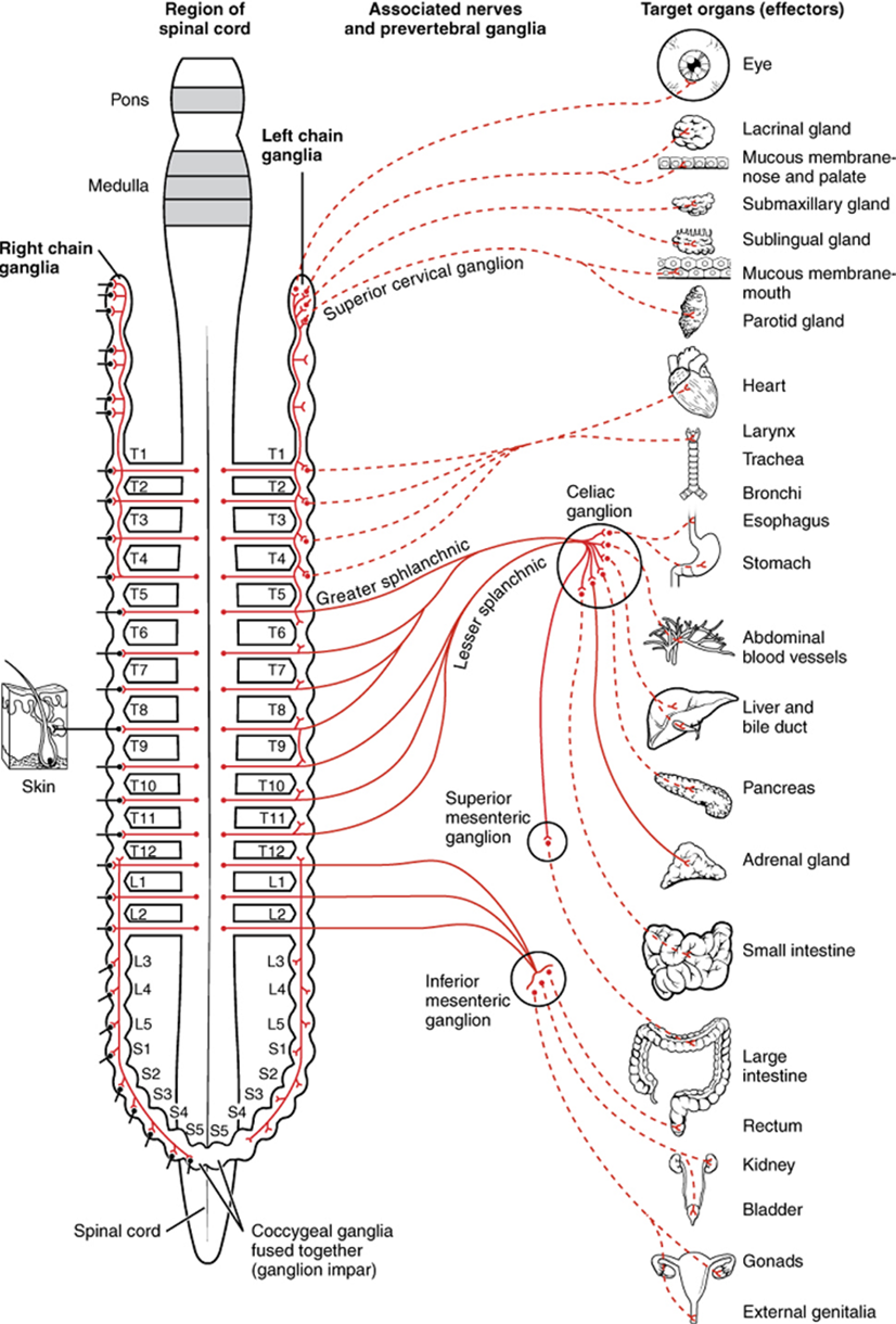
The projections of the sympathetic division of the autonomic nervous system diverge widely, resulting in a broad influence of the system throughout the body. As a response to a threat, the sympathetic system would increase heart rate and breathing rate and cause blood flow to the skeletal muscle to increase and blood flow to the digestive system to decrease. Sweat gland secretion should also increase as part of an integrated response. All of those physiological changes are going to be required to occur together to run away from the hunting lioness, or the modern equivalent. This divergence is seen in the branching patterns of preganglionic sympathetic neurons—a single preganglionic sympathetic neuron may have 10–20 targets. An axon that leaves a central neuron of the lateral horn in the thoracolumbar spinal cord will pass through the white ramus communicans and enter the sympathetic chain, where it will branch toward a variety of targets. At the level of the spinal cord at which the preganglionic sympathetic fibre exits the spinal cord, a branch will synapse on a neuron in the adjacent chain ganglion. Some branches will extend up or down to a different level of the chain ganglia. Other branches will pass through the chain ganglia and project through one of the splanchnic nerves to a collateral ganglion. Finally, some branches may project through the splanchnic nerves to the adrenal medulla. All of these branches mean that one preganglionic neuron can influence different regions of the sympathetic system very broadly, by acting on widely distributed organs.
Parasympathetic Division of the Autonomic Nervous System
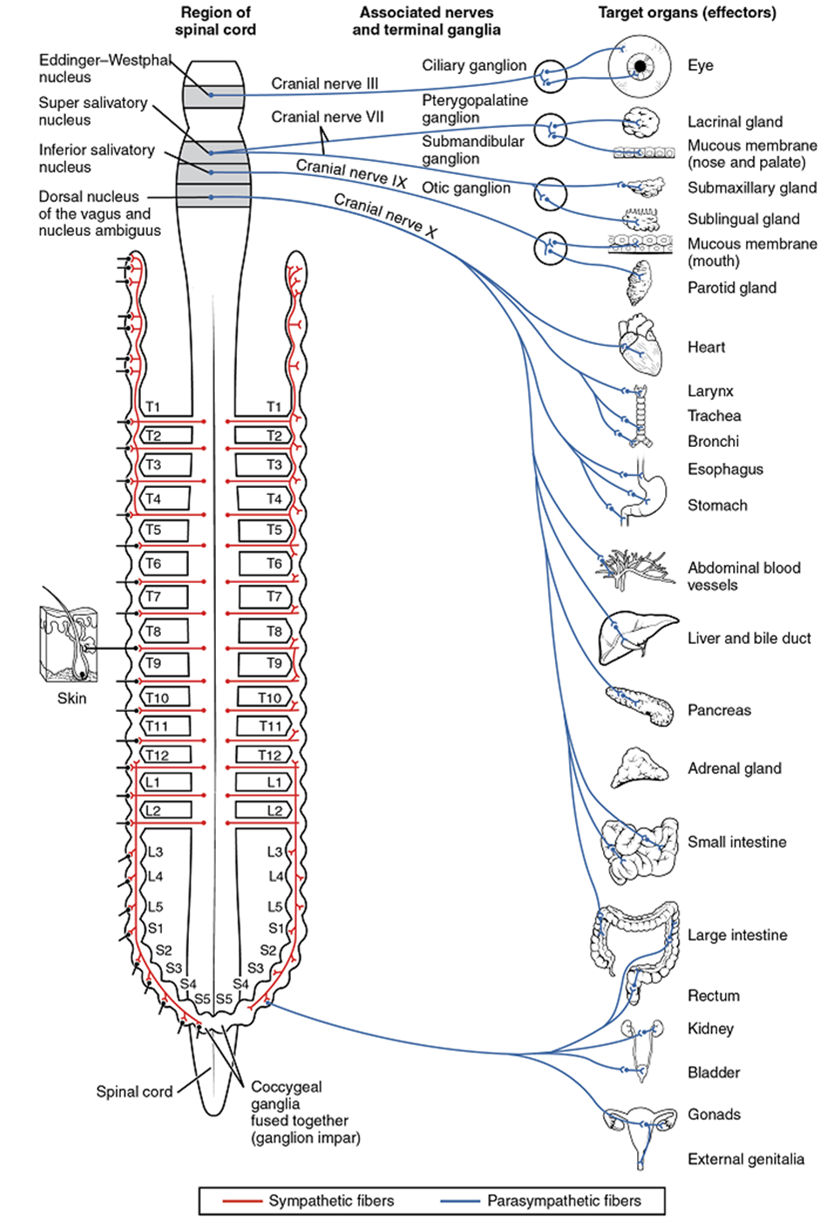
When not responding to an immediate threat, the parasympathetic system is generally more active than the sympathetic system. Many of the same effectors in the body are innervated by both divisions of the autonomic nervous system, but activation of each division tends to have opposing effects. Sympathetic system activation tends to increase activity in the respiratory, cardiovascular, and musculoskeletal systems while reducing activity in the digestive system. Parasympathetic system activation on the other hand tends to decrease activity in the respiratory, cardiovascular, and musculoskeletal systems while increasing activity in the digestive, and urinary systems. Reproductive systems are regulated by both sympathetic and parasympathetic innervation. Generally speaking, the activity of the many organs that receive input from both systems is dependent on whether neurons of the parasympathetic or sympathetic system are releasing more of their neurotransmitter onto each organ at a given time.
The parasympathetic division of the autonomic nervous system is named because its central neurons are located on either side of the thoracolumbar region of the spinal cord (para- = “beside” or “near”). The parasympathetic system can also be referred to as the craniosacral system (or outflow) because the preganglionic neurons are located in nuclei of the brain stem and the lateral horn of the sacral spinal cord.
The connections, or “circuits,” of the parasympathetic division are similar to the general layout of the sympathetic division with a few specific differences (Figure 5). The preganglionic fibres from the cranial region travel in cranial nerves, whereas preganglionic fibres from the sacral region travel in spinal nerves. The targets of these fibers are terminal ganglia, which are located near – or even within – the target organ. The postganglionic fibre projects from the terminal ganglia a short distance to the effector. These ganglia are often referred to as intramural ganglia when they are found within the walls target effector, or to the specific target tissue within the organ. Comparing the relative lengths of axons in the parasympathetic system, the preganglionic fibres are long and the postganglionic fibres are short because the ganglia are close to – and sometimes within – the target effectors.
Part 4: Receptor Pharmacology
Chemical Signaling in the Autonomic Nervous System
Where an autonomic neuron connects with a target, there is a synapse. The electrical signal of the action potential causes the release of a signaling molecule, which will bind to receptor proteins on the target cell. Synapses of the autonomic system are classified as either cholinergic, meaning that acetylcholine (ACh) is released, or adrenergic, meaning that norepinephrine is released. The terms cholinergic and adrenergic refer not only to the signaling molecule that is released but also to the class of receptors that each binds.
The cholinergic system includes two classes of receptor: the nicotinic receptor and the muscarinic receptor. Both receptor types bind to ACh and cause changes in the target cell. The nicotinic receptor is a ligand-gated cation channel and the muscarinic receptor is a G protein–coupled receptor. The receptors are named for, and differentiated by, other molecules that bind to them. Whereas nicotine will bind to the nicotinic receptor, and muscarine will bind to the muscarinic receptor, there is no cross-reactivity between the receptors. The situation is similar to locks and keys. Imagine two locks—one for a classroom and the other for an office—that are opened by two separate keys. The classroom key will not open the office door and the office key will not open the classroom door. This is similar to the specificity of nicotine and muscarine for their receptors. However, a master key can open multiple locks, such as a master key for the Biology Department that opens both the classroom and the office doors. This is similar to ACh that binds to both types of receptors. The molecules that define these receptors are not crucial—they are simply tools for researchers to use in the laboratory. These molecules are exogenous, meaning that they are made outside of the human body, so a researcher can use them without any confounding endogenous results (results caused by the molecules produced in the body).
The adrenergic system also has two types of receptors, named the alpha (α)-adrenergic receptor and beta (β)-adrenergic receptor. Unlike cholinergic receptors, these receptor types are not classified by which drugs can bind to them. All of them are G protein–coupled receptors. There are two types of α-adrenergic receptors, termed α1, and α2, and there are three types of β-adrenergic receptors, termed β1, β2 and β3. An additional aspect of the adrenergic system is that there is a second signaling molecule called epinephrine. The chemical difference between norepinephrine and epinephrine is the addition of a methyl group (CH3) in epinephrine. The prefix “nor-” actually refers to this chemical difference, in which a methyl group is missing.
The term adrenergic should remind you of the word adrenaline, which is associated with the fight-or-flight response described at the beginning of the chapter. Adrenaline and epinephrine are two names for the same molecule. The adrenal gland (in Latin, ad- = “on top of”; renal = “kidney”) secretes adrenaline. The ending “-ine” refers to the chemical being derived, or extracted, from the adrenal gland. A similar construction from Greek instead of Latin results in the word epinephrine (epi- = “above”; nephr- = “kidney”). In scientific usage, epinephrine is preferred in the United States, whereas adrenaline is preferred in Great Britain, because “adrenalin” was once a registered, proprietary drug name in the United States. Though the drug is no longer sold, the convention of referring to this molecule by the two different names persists. Similarly, norepinephrine and noradrenaline are two names for the same molecule.
All preganglionic fibres, both sympathetic and parasympathetic, release ACh. The postganglionic parasympathetic fibres also release ACh. Postganglionic sympathetic fibers release norepinephrine, except for fibers that project to sweat glands and to blood vessels associated with skeletal muscles, which release ACh.
Signaling molecules can belong to two broad groups. Neurotransmitters are released at synapses, whereas hormones are released into the bloodstream. These are simplistic definitions, but they can help to clarify this point. Acetylcholine can be considered a neurotransmitter because it is released by axons at synapses. The adrenergic system, however, presents a challenge. Postganglionic sympathetic fibres release norepinephrine, which can be considered a neurotransmitter. But the adrenal medulla releases epinephrine and norepinephrine into circulation, so they should be considered hormones.

Neural tissue associated with the digestive system that is responsible for nervous control through autonomic connections.
Localized collection of neuron cell bodies in the peripheral nervous system.
Sensory ganglion attached to the posterior nerve root of a spinal nerve.
Functional division of the nervous system that is responsible for homeostatic reflexes that coordinate control of cardiac and smooth muscle, as well as glandular tissue.
Cord-like bundle of axons located in the peripheral nervous system that transmits sensory input and response output to and from the central nervous system.
Bundle of axons in the central nervous system having the same function and point of origin
Type of tissue that serves to hold in place, connect, and integrate the body’s organs and systems.
One of twelve nerves connected to the brain that are responsible for sensory or motor functions of the head and neck.
Tenth cranial nerve; responsible for the autonomic control of organs in the thoracic and upper abdominal cavities.
Division of the anterior (ventral) cavity that houses the heart, lungs, esophagus, and trachea.
Single process of the neuron that carries an electrical signal (action potential) away from the cell body toward a target cell.
(In nervous system) a localized collection of neuron cell bodies that are functionally related; a “center” of neural function (plural= nuclei).
Functional division of the nervous system that is concerned with conscious perception, voluntary movement, and skeletal muscle reflexes.
Usually attached to bone, under voluntary control, each cell is a fiber that is multinucleated and striated.
Circuit of a reflex that involves a sensory input and motor output, or an afferent branch and an efferent branch, and an integrating center to connect the two branches.
Four muscles, that extend and stabilize the knee.
Change in voltage of a cell membrane in response to a stimulus that results in transmission of an electrical signal; unique to neurons and muscle fibres.
Muscle that opposes the action of an agonist.
Two-headed muscle that crosses the shoulder and elbow joints to flex the forearm while assisting in supinating it and flexing the arm at the shoulder.
Narrow junction across which a chemical signal passes from neuron to the next, initiating a new electrical signal in the target cell.
Three-headed muscle that extends the forearm.
Opposite side of the body.
Major region of the diencephalon that is responsible for relaying information between the cerebrum and the hindbrain, spinal cord, and periphery.
Outer gray matter covering the forebrain, marked by wrinkles and folds known as gyri and sulci.
One of six muscles originating out of the bones of the orbit and inserting into the surface of the eye which are responsible for moving the eye.
Branch of the autonomic nervous system associated with emergency systems ("fight of flight").
Division of the autonomic nervous system responsible for restful and digestive functions.
Organ that can cause a change in a value.
Alternate name for the sympathetic division of the autonomic nervous system that is based on the anatomical location of central neurons in the lateral horn of the thoracic and upper lumbar spinal cord.
Specifically referring to the cell body of a neuron in the autonomic system that is located in the central nervous system, specifically the lateral horn of the spinal cord or a brain stem nucleus.
Series of ganglia adjacent to the vertebral column that receive input from central sympathetic neurons.
Axon from a central neuron in the autonomic nervous system that projects to and synapses with a ganglionic neuron; sometimes referred to as a preganglionic neuron.
Axon from a ganglionic neuron in the autonomic nervous system that projects to and synapses with the target effector; sometimes referred to as a postganglionic neuron.
Inner layer of the adrenal glands that plays an important role in the stress response by producing epinephrine and norepinephrine.
Endocrine glands located at the top of each kidney that are important for the regulation of the stress response, blood pressure and blood volume, water homeostasis, and electrolyte levels.
Paired nerves that carry both autonomic and sensory fibres to the internal organs.
Chemical signal that is released from the synaptic end bulb of a neuron to cause a change in the target cell.
Alternate name for the parasympathetic division of the autonomic nervous system that is based on the anatomical location of central neurons in brain-stem nuclei and the lateral horn of the sacral spinal cord; also referred to as craniosacral outflow.
Protein molecule that contains a binding site for another specific molecule (called a ligand).
Synapse at which acetylcholine is released and binds to the nicotinic or muscarinic receptor.
An important neurotransmitter.
Synapse where norepinephrine is released, which binds to α- or β-adrenergic receptors.
Signaling molecule released as a neurotransmitter by most postganglionic sympathetic fibres as part of the sympathetic response, or as a hormone into the bloodstream from the adrenal medulla.
Signaling molecule released from the adrenal medulla into the bloodstream as part of the sympathetic response.
